Wednesday, 25 January 2012

Treatment of Orofacial Pain: TMD and Facial Neuraligias

Waking up in the morning with headaches? Painful popping in the jaw with chewing and yawning? Sharp, severe, lancinating pain in the head, face, ear, or jaw? You may be suffering from a type of orofacial pain!
Often the structure involved is the temporomandibular joint (TMJ) and the muscles surrounding this area. Pain may be due to a hypo- or hypermobile TMJ (too little or too much movement, respectively), derangement of the TMJ disc, or tightness of the muscles of mastication.
Your physiotherapist will take a detailed history of your current complaints, as well as a meticulous musculoskeletal assessment of your jaw and neck. We will be looking for asymmetry of facial structures, as well as movement deviations of the jaw through its available range of motion.

Picture taken from: http://www.primehealthchannel.com/tmj-disorder-anatomy-causes-symptoms-and-pain.html. Thank-you.

Another examination your physiotherapist will do will be checking the teeth for signs of bruxism (teeth clenching or grinding) as well as the insides of the cheek for damage. This may be evoked during sleep in the night or by stress during the day. Clients that have TMJ pain and are 'grinders' may benefit from having a night guard to help maintain the jaw in a slight rest position, and refrain from wearing their teeth down. 

On many occasions, temporary and relative rest of the jaw are required to improve healing. This may be as simple as temporarily eating softer foods, or cutting up the food into smaller pieces. Usually gum and pen chewing must cease and desist immediately.

Physiotherapy treatment of TMJ dysfunction (TMD) may joint mobilizations, stretching, myofascial release, acupuncture, and strengthening exercises. In a recent study conducted in Sweden (reviewing of systematic reviews and meta-analyses) found that there is evidence that night guards, acupuncture, exercises and postural retraining can be effective in alleviating TMD pain. Interestingly they found that electophysical agents (including ultrasound and laser) as well as surgery were ineffective, although could not be conclusive because of methodology variation between studies.

I would like to enforce that by no means is your physiotherapist the only professional to help you through your care of TMD. We will be working hand-in-hand with your dentist and possibly other health professionals such as your family physician, a neurologist, or an orofacial surgeon. Without treating the whole picture, we risk poor recovery.


List, T. & Axelsson, S. (2010). Management of TMD: evidence from systematic reviews and meta-analyses. Journal of Oral Rehabilitation. May 37, 430-451.

Friday, 20 January 2012

MRIs show: If you don't move it, you lose it!

I just happened to check out Discovery News Wednesday to see that there was a new article published in this month's Neurology Journal.

The study looked at 10 right hand dominant subjects that had sustained a right sided upper extremity injury that required immobilization. Each subject had an MRI within the first 48 hours of the injury, as well as one after 14 days of immobilization:

Exerpt: "The entire right arm was immobilized, preventing even small movements in the arm and hand."

Comparing the initial MRI to the 2 week follow-up, there was a decrease in the cortical thickness in the primary motor areas of the brain designating to the right arm and hand suggesting complete disuse of the limb will create plastic changes in the brain.

This image taken from: http://thoughtbroadcast.com/2011/02/10/thats-ok-i-didnt-need-that-brain-anyway/


Interestingly the area of the brain that controls motor function of the left upper extremity (the patient's non-dominant side) had increased in size, since most of these patients were required to now use the left arm for tasks such as feeding and brushing their teeth. Over the two weeks these patients became more dexterous with the left hand (objectified with a series of fine motor tests), and these changes can be seen objectively on MRI.

Although this study shows that extreme immobilization can result in plastic changes of the brain, one can apply this to patients who have been cast at a single joint. This also sparks the question that if you are generally inactive will that result in general loss of brain volume in motor areas?

The study by N. Langer, MSc and L. Jancke will also be following these patients after removal of the immobilization device. How long will it take for the cortical area to expand to pre-injury size, or will it achieve this thickness again?

N. Langer, et. al. (2012). Effects of limb immobilization on brain plasticity. Neurology. 78, 182-188.

Monday, 16 January 2012

This is why it is important to do your physio prescribed exercises!


No matter what the injury or condition, most if not all clients get at least one exercise. These are not just prescribed to take time out of your busy life, but they are meant to help control symptoms, and improve your recovery!

I'd like to bring to your attention an article in the Journal of Shoulder and Elbow Surgery concerning clients with tennis elbow (chronic lateral epicondylosis). The study took 21 patients that were categorized as having this chronic condition and separated them into an eccentric exercise group and into a physiotherapy no eccentric exercise group.

Eccentric training works extremely well for the rehabilitation for a number of conditions such as Achilles tendonosis. An eccentric muscle contraction occurs when the muscle being worked is lengthening under tension. This kind of exercise recruits more muscle fibers vs concentric muscle contractions (muscle shortening under tension). Eccentric muscle contractions can also be called the deceleration contraction or negatives in some gyms. Eccentric loading can take up to 40% more weight than concentric contractions allowing controlled tissue breakdown and more tissue remodeling.

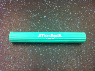
The eccentric exercise was done with a newer device that is a rubber bar. The rubber bar has a certain amount of elastic tension when deformed, and comes in a few different strengths. If you were to look to purchase these you can look online for the Cando Twist-N-Bend or the Thera-Band Flexbar. They range in price from $15-$30 depending on brand and tension.


Here is my flexbar below:


This image was taken from http://www.hygenicblog.com, the exercise was developed by physiotherapist Tim Tyler.


In both groups treatment was continued until their symptoms resolved or they had continued symptoms (about 8 weeks generally). The study found that self-reported functionality, point tenderness, and strength has significantly improved in the eccentric exercise group versus the therapy group without any eccentric training.


Unfortunately this study did not allow for the inclusion of a group that had both the physiotherapy and the eccentric training. Often with chronic tennis elbow, there can be myofascial trigger points in the extensor muscles of the wrist and elbow, and can often benefit from acupuncture to those points.

Tyler, T. F., et. al. (2010). Addition of isolated wrist extensor eccentric exercise to standard treatment for chronic lateral epicondylosis: A prospective randomized trial. Journal of Shoulder and Elbow Surgery, 19, 917-922.

Quick Ankle Update

Today will be the mark of the second week of rehabilitation of my ankle sprain, and things are going very smoothly! As you can see there are only mild sings of swelling in the front of my ankle, and no more blue-yellow bruising!


I have continued to work on my range of motion, especially end-range loading to help stretch out my scarred ligaments and ankle joint capsule. Elastic strengthening and wobble board training have also continued and progressed as now I am practicing single leg balance on the wobble board.

Knowing I cannot reach my ankle effectively and feel whether any of my joints have stiffened up, I decided to visit my physiotherapist who also happens to be the instructor of the last myofascial release course that I took in late 2011.


After an assessment, he could feel that my subtalar joint had limited and could benefit from manipulation. Since there was no discomfort with the pre-manipulative hold, my therapist applied a high velocity flick to the ankle, regaining lost mobility.


My physio also explained that often with an inversion sprain, that the peroneal nerve can become irritated by the quick stretch that is placed upon it by the motion of the ankle sprain. When this nerve is irritated it can make the muscles that evert (turn out) the foot tight. He suggested that I massage the outside of my leg at least once or twice a day, which I have followed up with as ordered.


The ankle looks and feels great, and I hope to be comfortable to run in the near future!

Monday, 9 January 2012

Day 6 - Post Ankle Sprain

It's the sixth day, and things are looking  and feeling great in my ankle. After the initial injury I was really thinking that the swelling would have been more widespread as well as the bruising.

I have been using the combination of acupuncture, ultrasound, as well as hot/cold applications to help reduce the inflammation as soon as possible. As you can see in the day 4 image below, the swelling on the lateral malleolus (the outside ankle bone) is not as dense as it once was. Initially there was at least 10mm of fluid, now we are taking about 1-2mm.


In these next images you can see the 6th day ankle, and the following is the application of ultrasound to the damaged area. If you read the comments of the last post, you can find more information concerning the application of ultrasound in physiotherapy.


As I noted previously, I wanted to do a little bit of a literature review of treatment of ankle sprains through physiotherapy and exercise therapy. The most applicable article that I could find that pertained to acute lateral ankle sprains was published in the Journal of Orthoapedic & Sports Physical Therapy (once again!).

The article is titled: Wobble Board Training After Partial Sprains of the Lateral Ligaments of the Ankle: A Prospective Randomized Study. The article took 48 subjects (each being in the athletic population) and were split into a training group and a no-training group (control). The training group completed a daily wobble board exercise program for 12 weeks, each session lasting about 15 minutes. Below you can see a little bit better the remaining swelling in the right ankle (this picture is taken into a mirror hence why it looks like the left ankle!).


The outcome measure was the subject's report of instability and re-injury. There was a significant difference between the two, concluding that those who did the wobble training effectively reduced the functional instability if the ankle (functional instability was stated to be the most common residual symptom following ankle sprains - ranging between 17-58% of patients, depending on the study).

I have been going on the wobble board daily, and look forward to being more stable on my feet!

Wester, J.N. et. al. (1996). Wobble Board Training After Partial Sprains of the Lateral Ligaments of the Ankle: A Prospective Randomized Study. Journal of Orthopaedic & Sports Physical Therapy, 23 (5), 332-336.


Friday, 6 January 2012

My Road to Recovery...

Wednesday morning, I decided to adjust the volume on the television in the adjacent gym space. The batteries for the remote have bitten the dust so I grabbed the flat bench to reach the manual buttons on the TV. Success! I could listen to my TSN sports highlights!

Without thinking I stepped off the bench only to find that my heel was placed on the caster to the bench and as my weight came down my ankle flipped inwards..

The snap was quite audible, like a branch breaking in a silent forest. My initial thoughts were concerned with how I was going to treat my patients today, and whether or not the ankle was broken. I walked around the gym several times to see if it would get better with the movement. Luckily I could weight-bear, this being a good indication that it is likely not fractured. 

I tossed ice on it immediately, though the outside aspect of my ankle was already beginning to swell and change colour. Below you can see the ankle at about 30 minutes after the injury.


My current working diagnosis on day 3 is a grade 2 inversion sprain with disruption of the ATF and CF ligaments as well as partial tearing of the lateral capsule. Although this does not sound that great, it is much better than a fracture or a grade 3 tear. The next image is my ankle at the end of day 2 (a little more colour and swelling, as I was standing most of the day).


Seeing as I have this blog up and running already, why not comment every so often about the recovery of my ankle sprain through physical therapy administered by myself.

Currently my treatment includes daily ultrasound over the swollen area and acupuncture. I am trying to ice the ankle 3x/day and will practice range of motion of the ankle constantly through drawing out the alphabet with my foot. Here you can see my ankle on day 3 with some of the needles in.


Today (day 3) I have added in strengthening of my ankle eversion muscles, as well as some wobble board exercises.

I will also be doing some research as to what treatments and exercises are shown to be the most effective as to be current with evidence based practice on lateral ankle sprains. Please check in for further updates!

Monday, 2 January 2012

For Women Dealing with Knee Pain!

A new randomized-control trial study has just been published in the Journal of Orthopaedic and Sports Physical Therapy concerning exercise intervention of females with patellofemoral pain (front knee pain).


This study had two groups: the first being prescribed an exercise intervention concentrating on hip abduction and hip external rotation strengthening, the other group being the no-exercise control.

The exercise program consisted of two elastic band strengthening exercises of the hip done 3x/week. After 8 weeks the exercise group showed a significant decrease in pain as well as function (based on self administered survey), and the control (non-exercise) group had no change in either outcomes.

This shows that although you may be dealing with pain in one area (the knee), the source of the problem may be coming from another location along the kinetic chain (in this case, the hip)!


Khayambashi, K. et al. (2012). The Effects of Isolated Hip Abductor and External Rotator Muscle Strengthening on Pain, Health Status, and Hip Strength in Females With Patellofemoral Pain: A Randomized Control Trial. The Journal of Orthopaedic and Sports Physical Therapy, 42(1), 22-29.